| |
 |
| Fig.
1 |
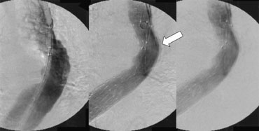
Digital
subtraction angiography during stent-grafting. Although
descending aortic aneurysm (Left) was excluded by the
stent grafts (Middle), a small amount of contrast medium
between the proximal stent graft and the native aorta
was detected (white arrow). Balloon compression reduced
this endoleak (Right). |
|
| |
 |
| Fig.
2 |
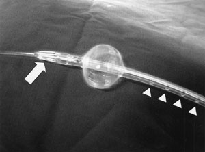
The
new stent-graft compression device. The white arrow
indicates a space for blood inflow. White arrow heads
indicate holes for blood outflow. |
|
| |
 |
| Fig.
3 |
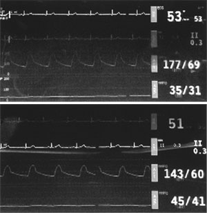
Hemodynamic
monitors during stent-graft compression using a conventional
aortic occlusion balloon (Upper) and the newly invented
stent-graft compression balloon (Lower). In each monitor,
electorocardiograms, blood pressure at the radial artery
and blood pressure at the left femoral artery are shown
from top to bottom. Systemic blood pressure at the radial
artery using a conventional balloon was higher than
using the new device, and mean blood pressure at the
left femoral artery using the new device was higher
than that using a conventional balloon. |
|
|


