Jpn. J. Vasc. Surg., 14: 151 - 158, 2005  
 
Fig. 1 Preoperative routine work for patients with critical ischemic limb.
 
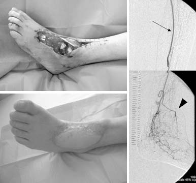
* Critical limb ischemia due to graft infection, DM: diabetes, HD: hemodialysis-dependent renal failure, Graft: bypass graft implanted previously, CFA: common femoral artery, SFA: superficial femoral artery, PFA: profound femoral artery, PABK: below knee popliteal artery, ATA: anterior tibial artery, PTA: posterior tibial artery, DP: dorsalis pedis artery, PTV: posterior tibial vein, MCF: musculocutaneous flap, MF: muscular flap
Table 1 Patient's characteristics and surgical procedures
 
Fig. 2 (a) Preoperative appearance of patient #3, showing extensive tissue loss around the foot joint with exposed bones and tendons. The limb was threatened by severe ischemia with infection because of repetitive bacterial emboli through infected artificial graft which had already removed. (b) Angiographic finding after latissimus dorsi flap transfer with femoro-posterior tibial artery bypass (arrow). Arrowhead indicates thoracodorsalis artery feeding flap. (c) Healed latissimus dorsi flap with salvaged foot photographed at one year after operation.
 
Fig. 3 56-year-old man with TAO, listed as patient #11. (a) Preoperative photograph of foot showing extensive tissue loss in forefoot with severe ischemia of remaining tissue. (b) Preoperative angiogram demonstrating only some collateral vessels with short diseased or recanalized segments of arteries, but no graftable segment of artery.
 
Fig. 4 Same patient shown in figure 3. (a) Scheme demonstrating surgical procedure of distal venous arterialization with free rectus flap transfer. Vein graft (VG) was anastomosed to posterior tibial vein whose valves had been destroyed surgically. Arrow indicates the intended direction of reversal flow. A: inferior epigastric artery feeding rectus flap. V: drainage vein of the flap, which anastomosed to another posterior tibial vein(*). (b) MRA finding 2 months after surgery. It is difficult to prove the retrograde flow through venous system of foot. However, MRA reveals early venous return (arrowheads). Arrow indicates rich microcirculation between flap and foot. (c) Postoperative appearance of successfully salvaged foot (5 months after surgery).
 
Table 2 The results of free flap transfer with bypass surgery
* Term to heal foot wound after free flap transfer, mo.: month