| |
 |
| Table |
Patients in whom partial endografting was used |
|
| |
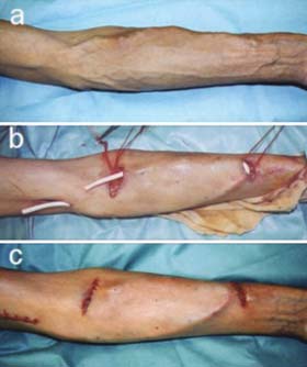 |
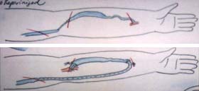
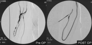
| Fig. 1 |
Surgical procedure. 1-a shows pre-operation, 1-b shows
endografting, 1-c shows post-operation. |
|
| |
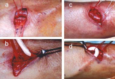 |
| Fig. 3 |
The prosthetic graft was inserted into the vein. |
|
| |
 |
| Fig. 4 |
Recoiling of the right antebrachial vein preoperatively and
underwent endografting using a 5-mm ePTFE graft in a 110-mm area of the antebrachial
vein. |
|
| |
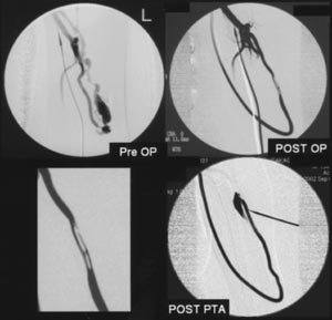 |
| Fig. 5 |
The patient with varication in the antebrachial vein and subsequent
thrombotic occlusion of the left antebrachial shunt. The graft was compressed
from outside by the puncture cannula. This area was repaired by using a balloon
catheter. |
|
| |
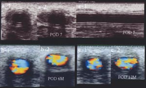 |
| Fig. 6 |
Ultrasound studies done 7 days, 6 months and 12 months after
the endografting procedure. a-1 and 3, b-1 and c-1 are the endograft area. a-2,
b-2 and c-2 are the nonendograft area. |
|
|