| |
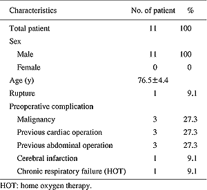 |
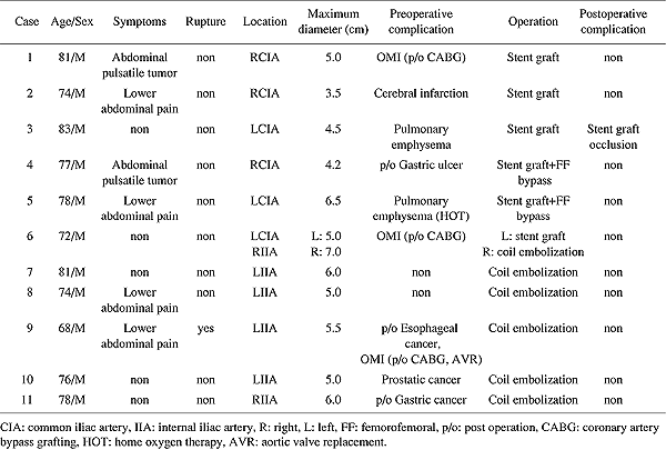
| Table
1 |
Clinical
characteristics |
|
| |
 |
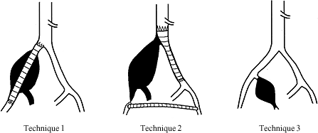
| Fig.
1 |
Schematic
figure of the operation.
A: Technique 1. SG was placed in the ipsilateral iliac
artery.
B: Technique 2. SG (aortouniiliac endografting) was
inserted distally from the abdominal aorta into the
contralateral iliac artery in conjunction with femoro-femoral
bypass.
C: Technique 3. Internal iliac artery coil embolization
was performed.
SG: stent graft. |
|
| |
 |
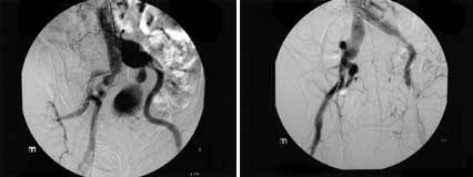
| Fig.
2 |
A:
Case 3. Preoperative angiography.
B: Case 3. Postoperative angiography (Fig. 1A). |
|
| |
 |
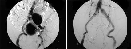
| Fig.
3 |
A:
Case 6. Preoperative angiography.
B: Case 6. Postoperative angiography (Fig. 1A, C). |
|
| |
 |
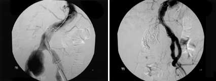
| Fig.
4 |
A:
Case 5. Preoperative angiography.
B: Case 5. Postoperative angiography (Fig. 1B). |
|
| |
 |
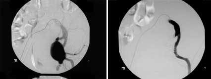
| Fig.
5 |
A:
Case 9. Preoperative angiography.
B: Case 9. Postoperative angiography (Fig. 1C). |
|
| |
 |
| Table
2 |
Summary
of isolated iliac artery aneurysm |
|


