| |

|
|
Fig. 1 2D DSA.
A small saccular shaped aneurysm was originated from the left radial artery (Arrow)
. Blood supply to the palmer artery arches was maintained only by the radial artery. |
|
| |
| |

|
|
Fig. 2 3D stereoview of the radial artery aneurysm.
A pair of 3D images were reconstructed in volume rending mode. |
|
| |
| |

|
|
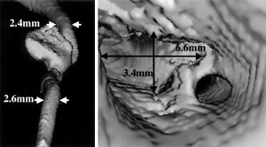
Fig. 3 Virtual endoscopy of radial artery aneurysm.
Left: volume rendering mode with three dimensional measurement of radial artery
diameters adjacent to the aneurysm. Right: A wide oval-shaped opening of the radial
artery was presented in the virtual endoscopic mode. |
|
| |
| |

|
|
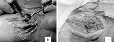
Fig. 4 Intraoperative photographs.
a: Before Resection.
b: After Reconstruction. |
|
| |
| |

|
|
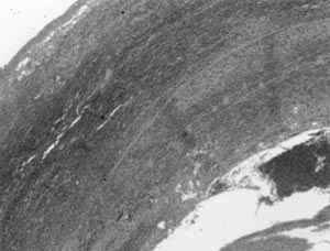
Fig. 5 Histology specimen from radial artery aneurysm.
Microscopic study of the resected specimen only showed fibrous tissue and organized
thrombus. (Hematoxylin & Eosin stain) |
|
| |
| |

|
|
Fig. 6 Post operative ultrasonography.
Ultrasonography was taken two months after operation.
a: B-mode image.
b: Power doppler image. |
|
| |
| |

|
|
| Table Three dimensional image devices. |
|
| |
|


